Research Highlights – Prof Giovanni Hearne
Home » Faculties of Science » Departments » Physics » Research » Research Highlights »Ultra-stability and enhanced stiffness of ~6nm TiO2 nanoanatase and eventual pressure-induced disorder on the nanometer scale
V. Pischedda, G. R. Hearne, A. M. Dawe, and J. E. Lowther
At 300 K ultrafine ~6 nm grain-size nanoanatase retains its structural integrity up to 18 GPa. There is progressive pressure-induced structural disorder to a highly disordered state at P>18 GPa. Signatures of short-range order persist to well beyond 18 GPa in both the synchrotron – x-ray diffraction and Raman data. Molecular dynamics simulations suggest disorder initiated in the surface shell of a nanograin with crystallinity being retained in the core. A bulk modulus B0 = 237±3 GPa for the nanoanatase (~30% higher than the bulk value) is derived from the P-V data, concordant with the MD calculations.
Physical Review Letters 96, 035509 (2006). (Also e-publication in Virtual Journal of Nanoscale Science & Technology 13, Issue 6 (2006)).
View Abstract and
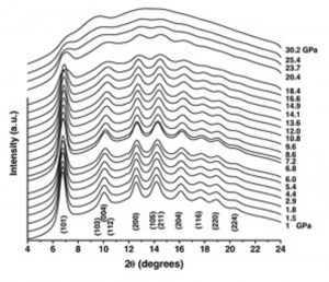
Figure. High-pressure powder x-ray diffraction spectra of nanoanatase up to 30 GPa at room temperature.
Thermally Induced Two-Step, Two-Site Incomplete 6A1<–>2T2 Crossover in a Mononuclear Iron(III) PhenolatePyridyl Schiff-Base Complex: A Rare Crystallographic Observation of the Coexistence of Pure S= 5/2 and 1/2 Metal Centers in the Asymmetric Unit
M.S. Shongwe, B.A. Al-Rashdi, H. Adams, M.J. Morris, M. Mikuriya, G.R. Hearne
The six-coordinate mononuclear iron(III) complexes [Fe(salpm)2]ClO4·0.5EtOH, [Fe(salpm)2]Cl, [Fe{(3,5-tBu2)-salpm}2]X (X=ClO4- or Cl-), and [Fe{(3,5-tBu2)-salpm}2]NO3·2H2O [Hsalpm = N-(pyridin-2-ylmethyl)- salicylideneamine; H(3,5-tBu2)-salpm = N-(pyridin-2-ylmethyl)-3,5-di-tert-butylsalicylideneamine] have been synthesized and isolated in crystalline form; their chemical identities have been ascertained by elemental analyses, FAB mass spectrometry, and infrared spectroscopy. The room-temperature effective magnetic moments [(8cMT)1/2 ~ 5.85−5.90 µB] of these complexes are consistent with the high-spin (S=5/2) ground state. These complexes are intensely colored on account of the strong pπ -> dπ* LMCT visible absorptions. Definitive evidence for the structures of [Fe(salpm)2]ClO4‚0.5EtOH and [Fe{(3,5-tBu2)-salpm}2]NO32H2O has been provided by single-crystal X-ray crystallography. The monomeric complex cations in both compounds comprise two uninegative phenolate−pyridyl tridentate Schiff-base ligands coordinated meridionally to the iron(III) to afford a distorted octahedral geometry with a trans,cis,cis-[FeO2N4] core. Whereas [Fe(salpm)2]ClO40.5EtOH undergoes a thermally induced 6A1 <-> 2T2 crossover, [Fe{(3,5-tBu2)-salpm}2]NO32H2O retains its spin state in the solid state down to 5 K. However, EPR spectroscopy reveals that the latter complex does exhibit a spin transformation in solution, albeit to a much lesser extent than does the former. The spin crossover in [Fe(salpm)2]ClO40.5EtOH has resulted in an unprecedented crystallographic observation of the coexistence of high-spin and low-spin iron(III) complex cations in equal proportions around 100 K. At room temperature, the two crystallographically distinct ferric centers are both high spin; however, one [Fe-(salpm)2]+ complex cation undergoes a complete spin transition over the temperature range ~200−100 K, whereas the other converts very nearly completely between 100 and 65 K; 10% of the complex cations in [Fe(salpm)2]-ClO40.5EtOH remain in the high-spin state down to 5 K.
Inorganic Chemistry 46, 9558-9568 (2007).
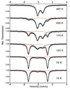
Figure. Mössbauer spectra of [Fe(salpm)2]ClO4·0.5EtOH.
Pressure-induced amorphization and a possible polyamorphism transition in nanosized TiO2: An X-ray Absorption Spectroscopy study
A.-M. Flank, P. Lagarde, J.-P. Itié, A. Polian , G.R. Hearne
The phenomenon of grain-size dependent pressure-induced amorphization (PIA) in TiO2 nanomaterials has been evidenced by several experiments in recent years. This has stemmed mainly from x-ray diffraction (XRD) and Raman studies of ultrafine grained anatase. Until now there is no experimental evidence of the length scale of disorder nor is there a clear picture of the amorphous structures, specifically in the case of pressure amorphised anatase-TiO2 starting material. The unresolved issues of the structural details and atomic-scale picture of the high-density amorphous (HDA) phase have now been addressed in an x-ray absorption spectroscopy (XAS) pressure study at the Ti K-edge. The local environment of the cation, to within a few nearest-neighbor shells, has been monitored up to ~30 GPa where the HDA phase is stabilized. In this phase the titanium atom is surrounded by 3±0.5 oxygens at 1.89 Å and 3±0.5 oxygens at 2.07 Å. The XAS results of this study suggest that a precursor ordered structural phase, different to that of anatase, is prevalent before amorphization occurs. The nature of this high-pressure stabilized precursor to amorphization likely depends on the starting experimental conditions at ambient pressure. In some cases this precursor has been identified as the columbite (a-PbO2-type) crystalline structure perhaps with only limited range order. Samples of this type appear to evidence a “memory effect” in that after cycling into the HDA phase (up to 30 GPa) where complete structural disorder prevails, this a-PbO2 structural intermediate is reestablished in a limited pressure range of the decompression cycle. It is also found that a new structure is stabilized in all cases of samples decompressed from the HDA phase to ambient conditions, characterized by fivefold coordinated Ti (2±0.5 oxygens at 1.84 Å and 2.5±0.5 oxygens at 2.06 Å) and is therefore structurally distinguishable from the HDA phase. These conceptual pictures are derived from the pressure dependence of both the extended x-ray-absorption fine structure (EXAFS) and the preedge parts of the absorption spectra.
Physical Review B 77, 224112 (2008).
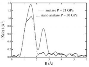
Figure. FT Magnitude of EXAFS of the HDA form of nanoanatase measured at 30 GPa, compared to the high-pressure form of bulk TiO2 at 21 GPa (i.e., baddeleyite-type phase).
Simplified manual fabrication of cubic-zirconia gem anvils for extended energy-range spectroscopic studies to routine high pressures of 100–150 kbar (10–15 GPa)
N.R. Jackson, R.M. Erasmus, and G.R. Hearne
Methodology has been developed so as to attain routine extreme conditions as high as 10–15 GPa in a gem anvil optical pressure cell using hand (manual) processed gem anvils. The anvils polished by a simplified hand held tool are inexpensive single crystal cubic zirconia (CZ) gems that have various optical advantages over diamond anvils. Appreciable pressures are attained with culet and corresponding sample cavity dimensions that are relatively convenient to load with sample material. Some technical details are provided as regards the simplified manual fabrication process, thus emphasizing the relative ease and cost effectiveness of the hand polishing technique for fabricating such high-pressure anvils. Raman spectroscopy measurements, in triple subtractive mode with confocal pinhole geometry, are used to exemplify the usefulness of the CZ gem anvil cell methodology in pressure tuning experiments. This is particularly convenient for conventional low wave-number (lattice mode regime). Raman high-pressure studies, which have not been reported previously in this context. Various other applications of such anvils are suggested.
Review of Scientific Instruments 81, 073903 (2010).
View Abstract
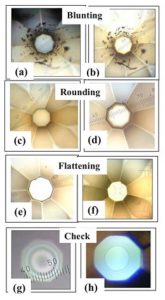
Figure. Polishing sequence to obtain final polished CZ gem anvil. (a) and (b) are the removal of sharp tip by polishing o a SiC abrading mat (P4000 grade); (c) and (d) are results of polishing on a soft mat impregnated with diamond powder spray to attain rounded culet edges. Darker shaded areas (in reflected light) at the perimeters are indicative of the curvature. Results of polishing on diamond grit impregnated polymer mat shown in (e) to obtain flattest possible truncated tip; (f) is further flattening and then viewed with an inverted microscope. Panel (g) is final quality-control test of anvil in (f) the appearance of a symmetric pattern of Newton’s rings as viewed through the table of the anvil in contact with a glass slide. Panel (h) shows typical picture of Newton’s ring pattern indicative of two well aligned anvils in a pressure cell with a thin glass slide sandwiched between them. Culet dimension in panels (e)–(h) has 380 mm diameter.
Pressure-induced quantum phase transition in Fe1−xCoxSi (x = 0.1, 0.2)
M.K. Forthaus, G.R. Hearne, N. Manyala, O. Heyer, R.A. Brand, D.I. Khomskii, T. Lorenz, and M.M. Abd-Elmeguid
We have investigated the effect of pressure on electrical transport and magnetic properties of ferromagnetic Fe1−xCoxSi alloys for x = 0.1 (TC ~ 11 K) and x = 0.2 (TC ~ 32 K) using electrical resistivity measurements up to about 30 GPa. We have also studied the magnetic properties of these samples (x = 0.1 and x = 0.2) and a sample with x = 0.3 (TC ~ 43 K) at ambient pressure using 57Fe Mossbauer-effect (ME) spectroscopy. The ME results indicate that the effective magnetic hyperfine field Beff at the 57Fe nucleus exhibits the same concentration dependence as the macroscopic magnetic moment and confirm that the onset of magnetic order is above x ∼ 0.02. The analysis of the high-pressure results reveals in both samples a gradual suppression of the ferromagnetic state to a quantum phase transition (QPT) at pressures of p ~ 11 GPa and p ~ 12 GPa for x = 0.1 and x = 0.2, respectively. High-pressure x-ray diffraction measurements on the three samples indicate very similar change of the volume of the cubic unit cell with pressure and exclude that the observed QPTs are connected with a structural phase transition. We discuss the observed instability of the ferromagnetic state with increasing Co concentration in the context of increasing local crystallographic disorder, which causes a change of the distribution of the helix wave vector as well as a modification of the ferromagnetic half-metallic state. We further show that, in the pressure-induced nonmagnetic metallic state, all samples, regardless of their different local crystallographic disorder, exhibit similar non-Fermi-liquid behavior [ρ(T)~T ]. Finally, we find a small but positive magnetoresistance in the high-pressure metallic state well beyond the QPT of Fe0.9Co0.1Si. This can be attributed to a slight field-induced modification of the spin majority and minority band, which leads to a very small magnetic moment.
Physical Review B 83, 085101 (2011). Highlighted as Editors’ Suggestion.
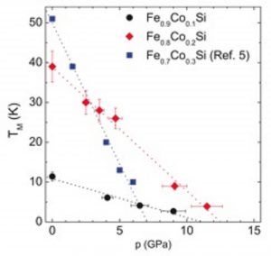
Figure. Pressure dependence of the upturn or kink at TM of the electrical resistivity at low temperatures, which is connected to the onset of magnetic order in Fe1−xCoxSi (x = 0.1, 0.2, 0.3). Dotted lines are linear fits to the experimental data points.
